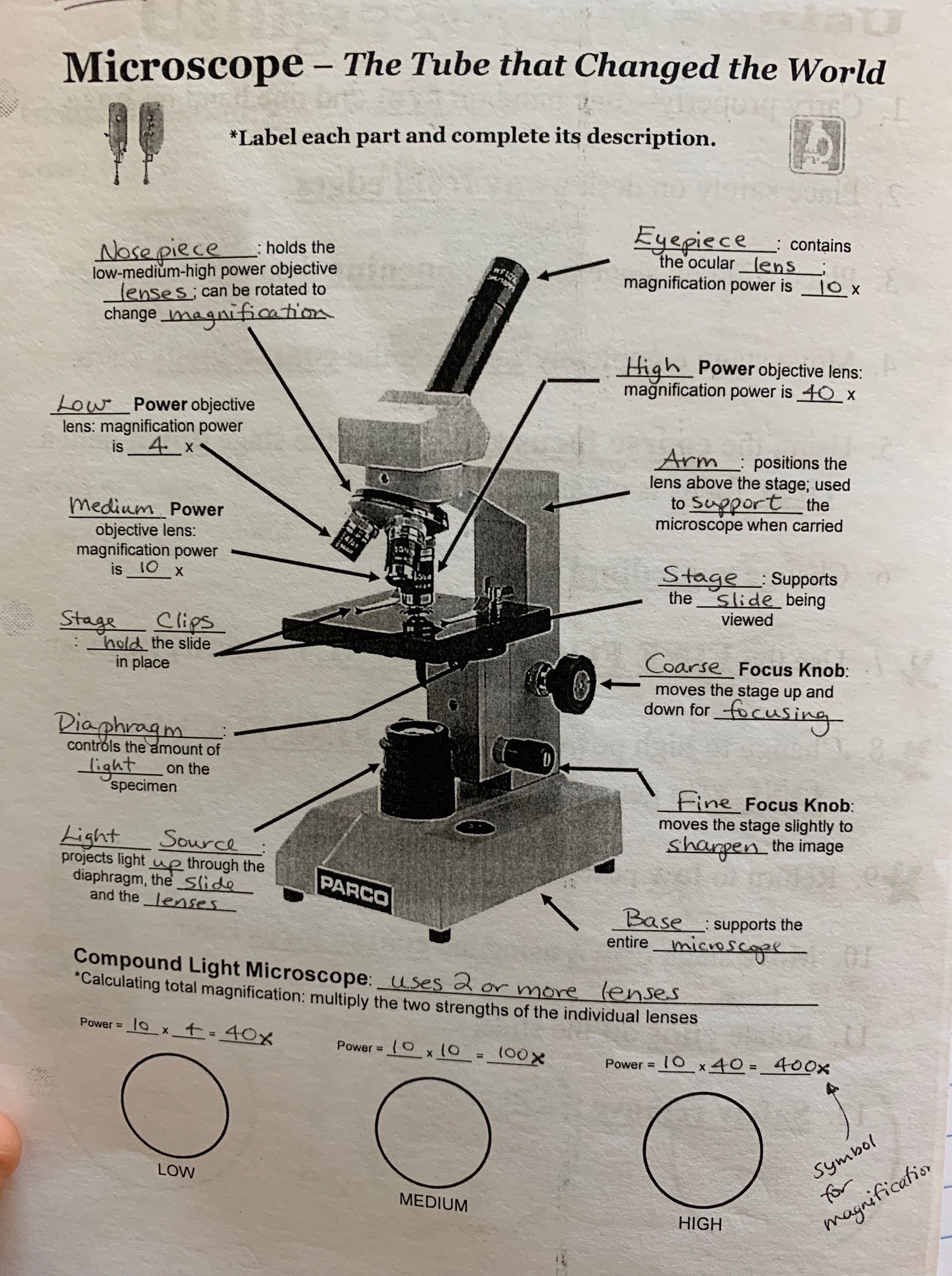
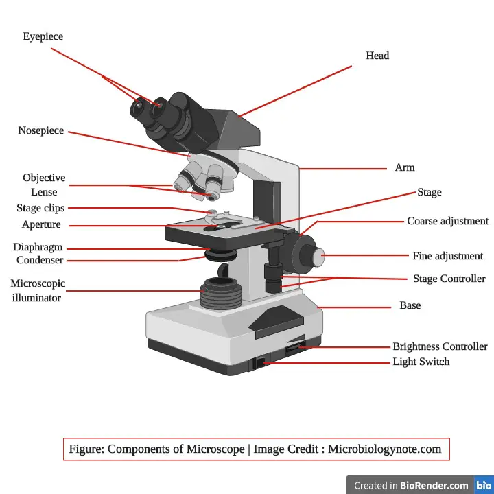
40 labels of a microscope and functions
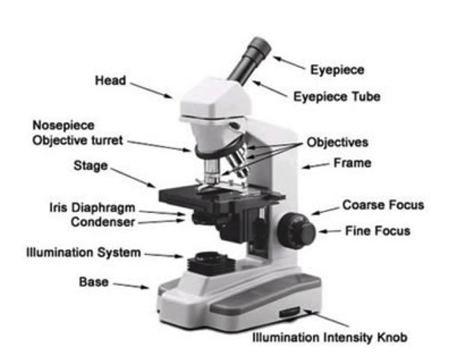
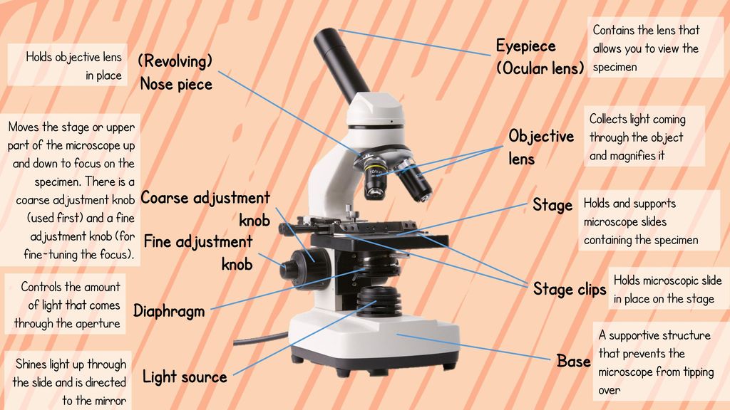
Parts of the Microscope with Labeling (also Free Printouts) Let us take a look at the different parts of microscopes and their respective functions. 1. Eyepiece it is the topmost part of the microscope. Through the eyepiece, you can visualize the object being studied. Its magnification capacity ranges between 10 and 15 times. 2. Body tube/Head It is the structure that connects the eyepiece to the lenses. Nervous System – Medical Terminology for Healthcare Professions The nervous system can be divided into regions that are responsible for sensation (sensory functions) and the response (motor functions), but there is a third function that needs to be included. Sensory input needs to be integrated with other sensations, as well as with memories, emotional state, or learning (cognition). Some regions of the nervous system are termed …
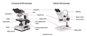
Microscope Parts, Function, & Labeled Diagram - slidingmotion Microscope parts labeled diagram gives us all the information about its parts and their position in the microscope. Microscope Parts Labeled Diagram The principle of the Microscope gives you an exact reason to use it. It works on the 3 principles. Magnification Resolving Power Numerical Aperture. Parts of Microscope Head Base Arm Eyepiece Lens
Labels of a microscope and functions
Compound Microscope Parts, Functions, and Labeled Diagram Compound Microscope Definitions for Labels. Eyepiece (ocular lens) with or without Pointer: The part that is looked through at the top of the compound microscope. Eyepieces typically have a magnification between 5x & 30x. Monocular or Binocular Head: Structural support that holds & connects the eyepieces to the objective lenses. Parts of Stereo Microscope (Dissecting microscope) – labeled … Compared to a compound microscope where the objectives attached to the nosepiece can be seen and identified individually (based on color bands and their respective labels), the objectives of a dissecting microscope are located in a cylindrical cone and, therefore, are not directly seen. For the stereo microscope that comes with multiple objective lens sets (fixed power style), the … Microscope: Parts Of A Microscope With Functions And Labeled Diagram. Functions of the optical components of a microscope. Examine, magnify, and produce a picture of a specimen on a slide using the microscope's optical devices. These are the constituent parts: Eyepiece:An alternative term for the eyepiece is the ocular. This includes the part of the microscope that allows for observation.
Labels of a microscope and functions. Conserved cell types with divergent features in human versus … 21.08.2019 · RNA-sequencing analysis of cells in the human cortex enabled identification of diverse cell types, revealing well-conserved architecture and homologous cell types as well as extensive differences ... Parts of a Compound Microscope and Their Functions - NotesHippo Arm: This is the portion of the microscope that connects the base to the head and the eyepiece tube to the base. It serves to support the microscope's head and is also used to transport the microscope. Stage: The stage is a flat and rectangular plate attached to the lower end of the arm. The specimen is placed on the stage so that the various ... 7 Types of Microscope and Their Functions - YaleTools A microscope is a laboratory tools that has a function to see micro-sized objects or organisms. This microscope is very common in laboratories because of its very important function. The principle of the microscope is to use two convex lenses, namely the objective lens and the eyepiece lens. Parts of a microscope with functions and labeled diagram - Microbe Notes Optical parts of a microscope and their functions The optical parts of the microscope are used to view, magnify, and produce an image from a specimen placed on a slide. These parts include: Eyepiece - also known as the ocular. This is the part used to look through the microscope. Its found at the top of the microscope.
Robert Hooke - Biography, Facts and Pictures Robert Hooke’s own illustration of his compound microscope, with labels added by this website. Hooke used his microscope to observe the smallest, previously hidden details of the natural world. His book Micrographia revealed and described his discoveries. Some people disputed his diagrams because they refused to believe what they showed. The world Hooke had discovered … Microscope Parts and Functions Flashcards | Quizlet Magnifies image 40X found on the nosepiece. Base. Support/bottom of the microscope, used to carry the microscope. Light Source. Provides light to enable us to see the specimen on the slide. Arm. Used in order to carry the microscope. Coarse Adjustment. moves the stage up or down a lot, used first when viewing the slide. ProtonMail Review 2022 - The Good and the Bad - RestorePrivacy Sep 05, 2022 · My email account was blocked instantly. Protonmail’s accounting system is a mess. They have an automated system that ruthlessly labels users as fraudulent regardless the fact that full payment was made within 24 hrs after a failed payment by paypal. The failure was caused by Protonmail’s accounting department. Parts of a Microscope Labeling Activity - Storyboard That Create a poster that labels the parts of a microscope and includes descriptions of what each part does. Click "Start Assignment". Use a landscape poster layout (large or small). Search for a diagram of a microscope. Using arrows and textables label each part of the microscope and describe its function. Copy This Storyboard* More options
Labeling the Parts of the Microscope | Microscope World Resources Labeling the Parts of the Microscope This activity has been designed for use in homes and schools. Each microscope layout (both blank and the version with answers) are available as PDF downloads. You can view a more in-depth review of each part of the microscope here. Download the Label the Parts of the Microscope PDF printable version here. Compound Microscope Parts - Labeled Diagram and their Functions Basically, compound microscopes generate magnified images through an aligned pair of the objective lens and the ocular lens. In contrast, "simple microscopes" have only one convex lens and function more like glass magnifiers. [In this figure] Two "antique" microscopes played significant roles in the history of biology. The Heart - Science Quiz - GeoGuessr The Heart - Science Quiz: Day after day, your heart beats about 100,000 times, pumping 2,000 gallons of blood through 60,000 miles of blood vessels. If one of your organs is working that hard, it makes sense to learn about how it functions! This science quiz game will help you identify the parts of the human heart with ease. Blood comes in through veins and exists via arteries—to control the ... Label Microscope Diagram - EnchantedLearning.com Using the terms listed below, label the microscope diagram. arm - this attaches the eyepiece and body tube to the base. base - this supports the microscope. body tube - the tube that supports the eyepiece. coarse focus adjustment - a knob that makes large adjustments to the focus. diaphragm - an adjustable opening under the stage, allowing ...
Monday 10/19/15 AIM: how do the parts of the compound light ...
Microscope Parts and Functions First, the purpose of a microscope is to magnify a small object or to magnify the fine details of a larger object in order to examine minute specimens that cannot be seen by the naked eye. Here are the important compound microscope parts... Eyepiece: The lens the viewer looks through to see the specimen.
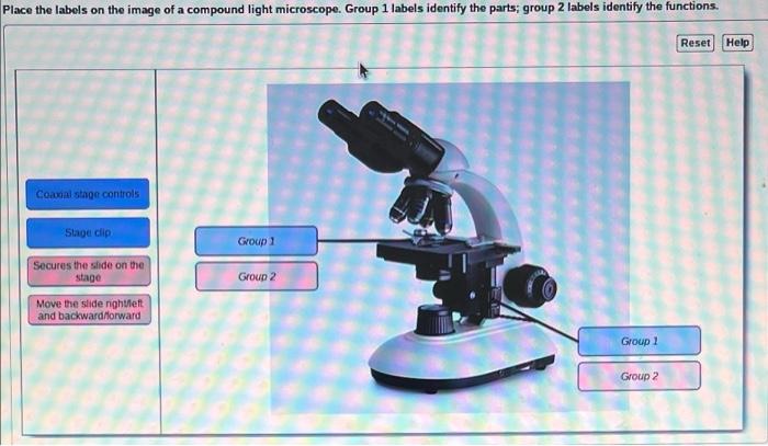
Solved Place the labels on the image of a compound light ...
Microscopy- History, Classification, Terms, Diagram - The Biology Notes History of Microscope. In the 1 st Century AD, the Romans invented the glass and used them to magnify objects.; In the early 14 th Century AD, eyeglasses were made by Italian spectacle makers.; In 1590, two Dutch spectacle makers, Hans, and Zacharias Jansen created the first microscope. It was a simple tube with 2 lenses system and had 9X magnification.

Compound Microscope: Parts of Compound Microscope
4.1: Cell Structure and Function - Medicine LibreTexts Aug 13, 2020 · A cell is the smallest living thing in the human organism, and all living structures in the human body are made of cells. There are hundreds of different types of cells in the human body, which vary in shape (e.g. round, flat, long and thin, short and thick) and size (e.g. small granule cells of the cerebellum in the brain (4 micrometers), up to the huge oocytes (eggs) produced in the female ...

Microscope labeled diagram
A Study of the Microscope and its Functions With a Labeled Diagram ... A Study of the Microscope and its Functions With a Labeled Diagram To better understand the structure and function of a microscope, we need to take a look at the labeled microscope diagrams of the compound and electron microscope. These diagrams clearly explain the functioning of the microscopes along with their respective parts.
microscope parts and functions - Quizizz
What are the label parts of the microscope? - Erasingdavid.com What are the functions of microscope? It is used in pedology (a study of soil particles) It is used by a dermatologist to find out various skin diseases. It is used in microbiology to study samples of algae,fungi etc. It is used by the jewellers to get a magnified view of the fine parts of the jewellery.

Simple Microscope Definition, Magnification, Parts And Uses
Microscope Parts & Functions - AmScope Microscope Parts and Functions Invented by a Dutch spectacle maker in the late 16th century, compound light microscopes use two sets of lenses to magnify images for study and observation. The first set of lenses are the oculars, or eyepieces, that the viewer looks into; the second set of lenses are the objectives, which are closest to the specimen.
Compound Microscope Parts, Functions, and Labeled Diagram ...
PharmaCircle This website uses cookies to help provide you with the best possible online experience. Please read our Terms & Conditions and Privacy Policy for information about ...

This is a common compound microscope. Label its parts from A ...
Simple Microscope - Parts, Functions, Diagram and Labelling Mirror - A simple microscope has a plano-convex mirror and its primary function is to focus the surrounding light on the object being examined. Lens - The biconvex lens is placed above the stage and its function is to magnify the size of the object being examined.

Compound Microscope Parts – Labeled Diagram and their ...
Parts of a Microscope - The Comprehensive Guide Step 1: Fully open field and condenser diaphragms and focus on specimen using x10 objective. Step 2: Fully close field diaphragm and adjust the condenser and focus so edges are as sharp as possible. Step 3: Use screws at front of condenser to centre field diaphragm and open field diaphragm to fill view. Step 4: Remove eyepiece and close down ...
Parts of a Microscope and Their Functions
List: Parts of a Microscope and their Function | Pathwooded Microscope Base The bottom piece of the microscope which provides support and stability for the microscope on your desk or tabletop is called the microscope base. Microscope Nosepiece Also known as the microscope turret, the revolving nosepiece of your microscope holds the different objective lenses just above the specimen you're examining.

Parts of Stereo Microscope (Dissecting microscope) – labeled ...
Microscope Types (with labeled diagrams) and Functions Simple microscope labeled diagram Simple microscope functions It is used in industrial applications like: Watchmakers to assemble watches Cloth industry to count the number of threads or fibers in a cloth Jewelers to examine the finer parts of jewelry Miniature artists to examine and build their work Also used to inspect finer details on products
Microscope Types (with labeled diagrams) and Functions
Microscope labeling and functions Flashcards | Quizlet Coarse Adjustment Knob Moves the body tube slightly to adjust the image; Fine Adjustment Knob Supports the body tube Arm Supports the slide being used Stage Projects or reflects light upward through the diaphragm Light Source Supports the microscope Base Body tube What is #1 called? Revolving nosepiece What is #2 called? Sets with similar terms

A diagram showing all of the parts of a compound light ...
Label the microscope — Science Learning Hub Use this interactive to identify and label the main parts of a microscope. Drag and drop the text labels onto the microscope diagram. eye piece lens diaphragm or iris coarse focus adjustment stage base fine focus adjustment light source high-power objective Download Exercise Tweet
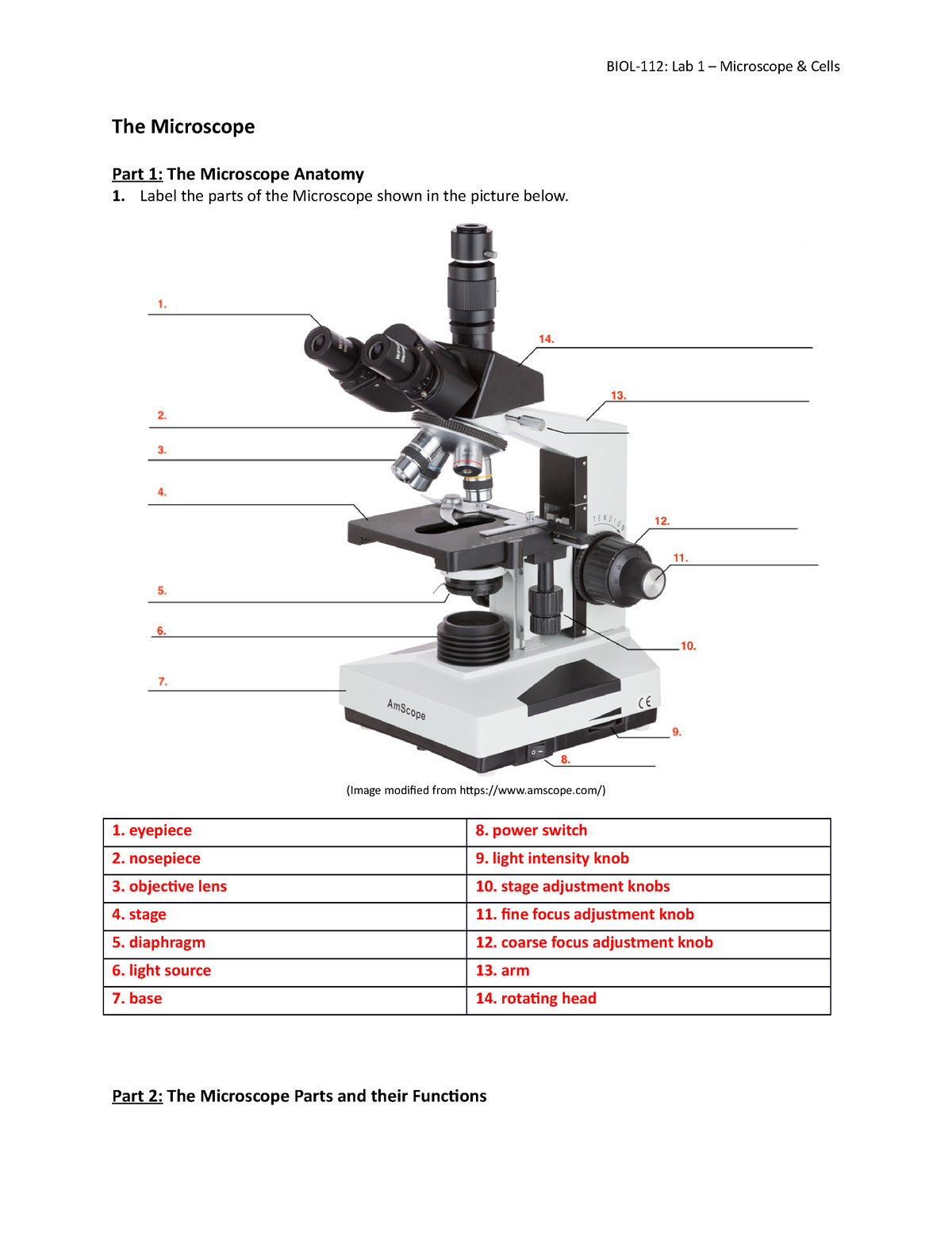
BIOL-112 Lab 1 Activtiy Worksheet Microscope & Cells - The ...
Autoclave - Wikipedia A thermal effluent decontamination system functions as a single-purpose autoclave designed for the sterilization of liquid waste and effluent. Air removal. It is very important to ensure that all of the trapped air is removed from the autoclave before activation, as trapped air is a very poor medium for achieving sterility. Steam at 134 °C (273 °F) can achieve a desired level of sterility …
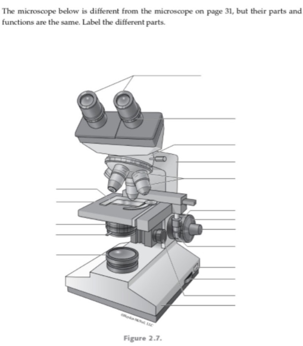
Solved The microscope below is different from the microscope ...
LAS X Industry Microscope software for Industry | Products Activate all relevant functions (e.g. for illumination settings, camera, measurements) with a few clicks ; Automatically store images on a regular basis with functionalities like autosave; Customizable user access. The software can handle multiple users who have different levels of microscope skills and diverse tasks to accomplish. Profiles according to user’s skills. The LAS …

Compound Microscope Parts, Function, & Diagram | What is a ...
PDF Label parts of the Microscope Label parts of the Microscope: . Created Date: 20150715115425Z

Compound Microscope Parts – Labeled Diagram and their ...
Microscope parts — Science Learning Hub In this activity, students identify and label the main parts of a microscope and describe their function. By the end of this activity, students should be able to: identify the main parts of a microscope. describe the function of the different parts of a microscope. Download the Word file (see link below) for: background information for teachers.
Compound Microscope Parts, Functions, and Labeled Diagram ...
Microscope, Microscope Parts, Labeled Diagram, and Functions Microscopes magnify or enlarge small objects such as cells, microbes, bacteria, viruses, microorganisms etc. at a viewable scale for examination and analysis. Microscopes consist of one or more magnification lenses to enlarge the image of the microscopic objects placed in the focal plane.

Microscope Parts and Functions
22 Parts Of a Microscope With Their Function And Labeled Diagram Invented by a Dutch spectacle maker in the late 16th century, light microscopes use lenses and light to magnify images. Generally a microscope works on the basis of resolution and magnification. The magnifying power of a microscope is an expression of the number of times the object being examined appears to be enlarged and is a dimensionless ratio.
Parts of Stereo Microscope (Dissecting microscope) – labeled ...
Microscope: Parts Of A Microscope With Functions And Labeled Diagram. Functions of the optical components of a microscope. Examine, magnify, and produce a picture of a specimen on a slide using the microscope's optical devices. These are the constituent parts: Eyepiece:An alternative term for the eyepiece is the ocular. This includes the part of the microscope that allows for observation.

Label the microscope — Science Learning Hub
Parts of Stereo Microscope (Dissecting microscope) – labeled … Compared to a compound microscope where the objectives attached to the nosepiece can be seen and identified individually (based on color bands and their respective labels), the objectives of a dissecting microscope are located in a cylindrical cone and, therefore, are not directly seen. For the stereo microscope that comes with multiple objective lens sets (fixed power style), the …
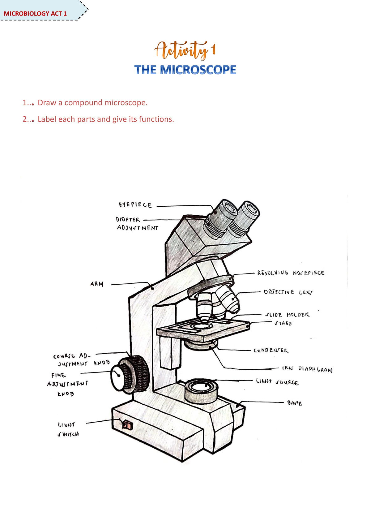
Microscope Activity - MICROBIOLOGY - 1... Draw a compound ...
Compound Microscope Parts, Functions, and Labeled Diagram Compound Microscope Definitions for Labels. Eyepiece (ocular lens) with or without Pointer: The part that is looked through at the top of the compound microscope. Eyepieces typically have a magnification between 5x & 30x. Monocular or Binocular Head: Structural support that holds & connects the eyepieces to the objective lenses.

Labeling the Parts of the Microscope | Microscope activity ...

List: Parts of a Microscope and their Function | Pathwooded

Label Microscope Diagram - EnchantedLearning.com

Parts of a Microscope Labeling Activity

Labeling a Microscope Free Worksheet Pack

Compound Microscope- Definition, Labeled Diagram, Principle ...

This is a common compound microscope. What the labelling D ...

Parts of the microscope activity

Simple Microscope - Parts, Functions, Diagram and Labelling ...
Parts and Functions of a Microscope - ppt download

2.2 Molecular make up of cells | Cells: the basic units of ...
(159).jpg)
Microscope Quiz: How Much You Know About Microscope Parts And ...
Microscope Parts and Functions

13 parts of the Compound Light Microscope Diagram | Quizlet

Pin on Micellaneous

Label the microscope — Science Learning Hub

Compound Microscope Parts – Labeled Diagram and their ...
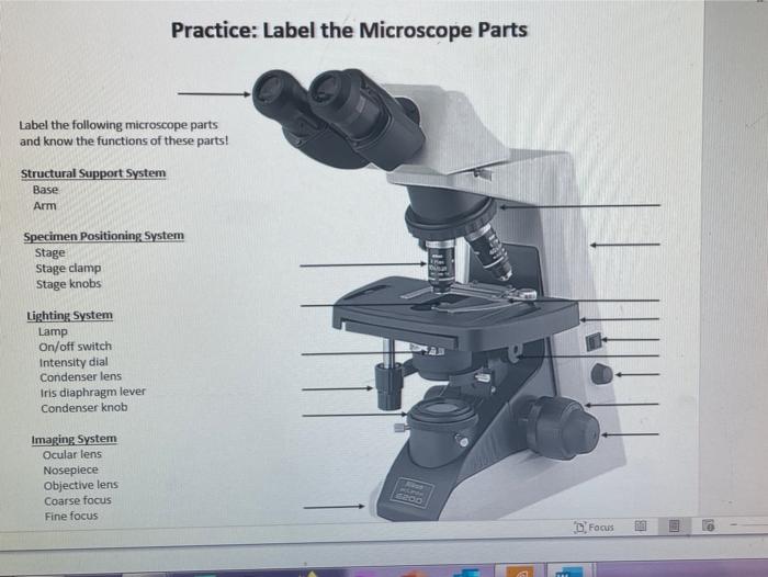
Solved Practice: Label the Microscope Parts Label the | Chegg.com
Post a Comment for "40 labels of a microscope and functions"