38 microscope with labels and functions
LAS X Industry Microscope software for Industry | Products ... Measure parameters, such as the length, area, diameter, angle, or perimeter of objects you mark with adjustable tracing lines, drawing directly in the live images. Add labels for easy analysis. Apply measurements to several images to determine statistical trend and compare data in measurement templates. Parts of a Microscope - The Comprehensive Guide Step 1: Fully open field and condenser diaphragms and focus on specimen using x10 objective. Step 2: Fully close field diaphragm and adjust the condenser and focus so edges are as sharp as possible. Step 3: Use screws at front of condenser to centre field diaphragm and open field diaphragm to fill view. Step 4: Remove eyepiece and close down ...
Microscope Parts, Function, & Labeled Diagram - slidingmotion Microscope parts labeled diagram gives us all the information about its parts and their position in the microscope. Microscope Parts Labeled Diagram The principle of the Microscope gives you an exact reason to use it. It works on the 3 principles. Magnification Resolving Power Numerical Aperture. Parts of Microscope Head Base Arm Eyepiece Lens

Microscope with labels and functions
Parts of the Microscope with Labeling (also Free Printouts) Let us take a look at the different parts of microscopes and their respective functions. 1. Eyepiece it is the topmost part of the microscope. Through the eyepiece, you can visualize the object being studied. Its magnification capacity ranges between 10 and 15 times. 2. Body tube/Head It is the structure that connects the eyepiece to the lenses. Electron microscope - Wikipedia An electron microscope is a microscope that uses a beam of accelerated electrons as a source of illumination. As the wavelength of an electron can be up to 100,000 times shorter than that of visible light photons , electron microscopes have a higher resolving power than light microscopes and can reveal the structure of smaller objects. Light Microscope: Functions, Parts and How to Use It The function of the light microscope is based on its ability to focus a beam of light through a very small and transparent specimen, to produce an image. The image is then passed through one or two lenses for magnification to view. The transparency of the specimen allows for easy and fast light penetration.
Microscope with labels and functions. ch 8 mastering biology Flashcards | Quizlet Looking through a light microscope at a dividing cell, you see two separate groups of chromosomes on opposite ends of the cell. New nuclear envelopes are taking shape around each group. The chromosomes then begin to disappear as they unwind. Microscope Parts & Functions - AmScope Microscope Parts and Functions Invented by a Dutch spectacle maker in the late 16th century, compound light microscopes use two sets of lenses to magnify images for study and observation. The first set of lenses are the oculars, or eyepieces, that the viewer looks into; the second set of lenses are the objectives, which are closest to the specimen. Introduction to three-dimensional image processing skimage.exposure contains a number of functions for adjusting image contrast. These functions operate on pixel values. Generally, image dimensionality or pixel spacing does not need to be considered. Gamma correction, also known as Power Law Transform, brightens or darkens an image. The function \(O = I^\gamma\) is applied to each pixel in the ... Microscope labeling and functions Flashcards | Quizlet Microscope labeling and functions STUDY Flashcards Learn Write Spell Test PLAY Match Gravity Created by mveet Terms in this set (27) Separates the eyepiece lens from the objective lenses Body Tube Holds the low-power and high-power objective lenses; allows the lenses to rotate for viewing Revolving Nosepiece Magnifies about 4x
Simple Microscope - Parts, Functions, Diagram and Labelling Stereo microscope/dissecting microscope - It can magnify objects by up to 300 times. It is used to visualize opaque objects that cannot be visualized using a compound microscope. Confocal microscope - It uses laser light to scan a dyed sample. Scanning electron microscope - Instead of light, this type of microscope uses electron. 5 Types of Microscopes with Definitions, Principle, Uses, Labeled Diagrams Parts of a microscope with functions and labeled diagram; Electron Microscope. It was invented by Ernst Ruska in 1931. It differs from a light microscope in various ways. There are two types of EM: Transmission EM (MC type, examine the internal structure, resolution 0.5 nm, gives 2-dimensional view) What is label in microscope? - Gowanusballroom.com Use this with the Microscope parts activity to help students identify and label the main parts of a microscope and then describe their functions. Drag and drop the text labels onto the microscope diagram. What is the body tube part of a microscope? It is the structure that connects the eyepiece to the lenses. Image 2: The body tube part of a ... Microscope With Labeled Parts and Functions - 24 Hours Of Biology Optical parts and the functions The optical parts of the microscope are used to view, enlarge, and produce an image from a sample placed on a slide. These parts include Eyepiece: Eyepiece also contains ocular lens. It enhance the image of the viewer. This part is used for checking through the microscope. Eyepiece is found at the upper part of it.
Confocal Microscopy - an overview | ScienceDirect Topics A confocal microscope was invented in 1951 by Marvin Minsky, a postdoctoral fellow at Harvard University studying neural networks in living brain (Minsky, 1988).In 1957, Minsky patented the concept of confocal imaging, the illumination and detection of a single diffraction-limited spot in a specimen (Fig. 1A). Microscope, Microscope Parts, Labeled Diagram, and Functions Illuminator: Illuminator is the most important microscope parts and it serve as light source for a microscope during slide specimen visualization. It is a continuous source of light (110 volts) used in place of a mirror. The mirror of microscope is used to reflect light from the external light source up through the bottom of the stage. Microscope: Types of Microscope, Parts, Uses, Diagram - Embibe There microscope anatomy includes three structural parts, i.e. head, base, and arm. Head - This is also known as the body; it carries the optical parts in the upper part of the microscope.. Base - It acts as microscopes support.It also carries microscopic illuminators. Arms - The microscope arm connects the base and the head and the eyepiece tube to the microscope base. Microscopy- History, Classification, Terms, Diagram - The Biology Notes History of Microscope. In the 1 st Century AD, the Romans invented the glass and used them to magnify objects. In the early 14 th Century AD, eyeglasses were made by Italian spectacle makers. In 1590, two Dutch spectacle makers, Hans, and Zacharias Jansen created the first microscope. It was a simple tube with 2 lenses system and had 9X ...
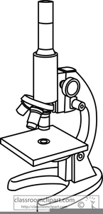
Compound Microscope Clipart | Free Images at Clker.com - vector clip art online, royalty free ...
Parts of a Microscope Labeling Activity - Storyboard That Create a poster that labels the parts of a microscope and includes descriptions of what each part does. Click "Start Assignment". Use a landscape poster layout (large or small). Search for a diagram of a microscope. Using arrows and textables label each part of the microscope and describe its function.

34 Label Microscope - Labels Design Ideas 2020
Microscope Labeling Worksheets & Teaching Resources | TpT This resource contains 1 worksheet for students to label the parts of a microscope and 1 worksheet for students to complete a chart detailing the functions of each microscope part. Answer key included. Resource comes as both a PDF for printing and a Microsoft Word document for editing or for a digi
Molecular Expressions: Science, Optics & You - Olympus MIC-D: Brightfield Gallery - Spiderwort ...
Microscope Types (with labeled diagrams) and Functions Simple microscope labeled diagram Simple microscope functions It is used in industrial applications like: Watchmakers to assemble watches Cloth industry to count the number of threads or fibers in a cloth Jewelers to examine the finer parts of jewelry Miniature artists to examine and build their work Also used to inspect finer details on products

Simple Microscope Labeled Diagram - Micropedia
Types of Microscopes: Definition, Working Principle, Diagram ... - BYJUS There are also microscope types that find application in metallurgy and studying three-dimensional samples. In this article, there are 5 such microscope types that are discussed along with their diagram, working principle and applications. These five types of microscopes are: Simple microscope. Compound microscope.

31 Drag The Label To The Appropriate Part Of The Microscope. - Labels Design Ideas 2020
Parts of a microscope with functions and labeled diagram - Microbe Notes Microscopes are instruments that are used in science laboratories to visualize very minute objects such as cells, and microorganisms, giving a contrasting image that is magnified. Microscopes are made up of lenses for magnification, each with its own magnification powers.
Post a Comment for "38 microscope with labels and functions"